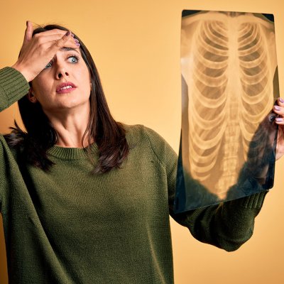
Recently, I had a young, otherwise healthy, male patient (we’ll call him Fred) in his mid-40’s who presented with low back pain. This is quite common in my small Idaho town as the economy is driven primarily by blue-collar jobs like farming and factory work. Patients like Fred come into the clinic every single day. Fred was very active and spent most mornings working out at the local gym, squatting and deadlifting. He sought medical care when he started to experience pain during weight-lifting activities. His physician ordered an MRI and the patient was diagnosed with “Degenerative Disc Disease.” Fred stated that no other information aside from his diagnosis was provided to him following his MRI.
During his initial evaluation with me, Fred expressed fear of continuing to lift weights because continued lifting would cause his spine to “crumble” and “break.” During my initial visit with Fred, I was able to get a copy of his MRI report. It found “mild degenerative changes in the lumbar spine.” I showed Fred what the report actually said, emphasizing the word “mild.” I next explained how common these findings are in men of his age, as well as how MRI findings are poorly correlated with low back pain symptoms (Suri, Kasch). Fred immediately expressed relief to me as he had been worried about never being able to do the things he enjoys ever again for fear of making his back worse. I met Fred at the local gym my clinic is affiliated with for our next visit. While he did still have some painful symptoms, his anxiety surrounding them was greatly reduced. We spent the visit going through regressions for common lifts that he could do without aggravating his symptoms. We also discussed how to progress from these back into his regular routine.
This is a clear example of how imaging (and the jargon we use regarding these images) can negatively impact how a patient experiences pain. The past decade has produced several studies that show an early MRI can actually produce worse outcomes in terms of healthcare costs and utilization, but also in overall disability and pain7,8. In short, early use of MRI imaging (except in the presence of red flags) is likely to make patients feel worse.
As physical therapists, we are unlikely to affect how physicians use advanced imaging with their patients. In my practice, patients have typically already undergone an MRI by the time they walk through my door. They typically enter the clinic fearful about a new diagnosis and worried their condition will devolve into a lifetime of pain and disability, as every patient knows someone who has dealt with debilitating back pain. Physical therapists can play an important role in contextualizing imaging findings for patients to help ease their anxiety and avoid iatrogenic pain.
Patients need to understand how common abnormal lumbar spine MRI findings are, even in asymptomatic individuals. A systematic review by Brinjikji et al noted that even people in their 20’s exhibited “abnormal” MRI findings about a third of the time. As people age, the odds of detecting abnormalities increases to up to 96% in those over 802. While it may seem paradoxical, these findings are quite common, even in people without pain. As our patients start to understand this (and it may take more than one visit), the anxiety surrounding their pain will drop significantly over time. It may take a little more time for their pain to resolve completely, but their experience with pain will be much improved, as they exhibit less kinesiophobia and fear-avoidance behaviors. This will allow the patient to enjoy favored activities, even in the presence of pain.
If we are lucky enough to see patients before they go in for imaging, we have an excellent opportunity to prime patients for what they should expect to see based on their demographic information, as well as on the findings of our physical examination of the patient. For example, another patient I recently evaluated (let’s call him Bill) presented with right shoulder pain subsequent to a workplace injury. Aside from pain and tenderness throughout the right shoulder girdle and the right side of his neck, the physical examination was unremarkable with no deficits in strength or range of motion. Bill said his physician had scheduled an MRI the following week.
Bill is a male in his mid-50’s who has made a good living working a job that requires lots of manual labor. During the visit, he had expressed concern that he had torn his rotator cuff and that this would negatively affect his ability to return to full duty at a job he loved. I took the time to help him understand that his MRI was likely to show some sort of rotator cuff pathology based on his age, gender, and history of manual labor. In spite of these potential findings, I told Bill that his shoulder was strong and stable based on my exam findings, and that “abnormal” findings were very common, even in asymptomatic individuals3. As Bill gets ready to receive the results of his MRI, he can rest easy knowing he doesn’t have to jump to surgery if his images show an abnormality that probably isn’t related to his current pain.
Whether we are meeting with a patient before or after their imaging, PTs should play a leading role in helping patients understand what is and isn’t normal. Many of the “degenerative” findings seen on advanced images simply reflect the realities of normal aging for most individuals and are likely unrelated to their pain2. Only if the patient's clinical presentation matches with the imaging results should we be emphasizing the scans at all. Oftentimes, if the imaging results show degenerative changes, but the patient’s symptoms don’t match their scans, I will refer to these degenerative changes simply as “normal, age-related changes.”
Minimizing our own noceboic language will also help ease patient’s anxieties surrounding advanced imaging. Nocebo (the opposite of placebo), refers to a phenomenon where negative expectations yield more negative results than would otherwise be expected. The words we use when we talk with our patients can have a profound impact on how our patients experience their pain as we temper expectations about the severity of their imaging/pathology. I frequently refer to a recent JOSPT viewpoint article5 as it provides a useful table of potentially harmful terminology and good replacements to use to minimize the noceboic effect they have on patients.
Surprisingly, abnormal findings on MRI are completely normal. As physical therapists, we have the opportunity to serve as a bridge between our patients and their understanding of these images. In tandem with a thorough physical examination, we can help patients contextualize their imaging findings and help break them out of a cycle of fear-avoidance and anxiety. These conversations must be had, and physical therapists are in an excellent position to have them. If we take advantage of this opportunity, I’ve no doubt we will further advance our vision of transforming society and improving the human experience1.
References
- American Physical Therapy Association. Vision, mission, and strategic plan. APTA. https://www.apta.org/apta-and-you/leadership-and-governance/vision-mission-and-strategic-plan. Accessed November 7, 2022.
- Brinjikji W, Luetmer PH, Comstock B, et al. Systematic literature review of imaging features of spinal degeneration in asymptomatic populations. American Journal of Neuroradiology. 2014;36(4):811-816.
- Girish G, Lobo LG, Jacobson JA, Morag Y, Miller B, Jamadar DA. Ultrasound of the shoulder: Asymptomatic findings in men. American Journal of Roentgenology. 2011;197(4).
- Kasch, Richard MDa; Truthmann, Julia PhDb; Hancock, Mark J. PhDc; Maher, Christopher G. DMedScd; Otto, Markus PhDe; Nell, Christopher MDe; Reichwein, Niklasa; Bülow, Robin MD, MSce; Chenot, Jean-François MD, MPHb; Hofer, Andre MDa; Wassilew, Georgi MDa; Schmidt, Carsten Oliver PhDf. Association of Lumbar MRI Findings with Current and Future Back Pain in a Population-based Cohort Study. SPINE 47(3):p 201-211, February 01, 2022.
- Stewart M, Loftus S. Sticks and stones: The impact of language in musculoskeletal rehabilitation. Journal of Orthopaedic & Sports Physical Therapy. 2018;48(7):519-522.
- Suri, P., Boyko, E.J., Goldberg, J. et al. Longitudinal associations between incident lumbar spine MRI findings and chronic low back pain or radicular symptoms: retrospective analysis of data from the longitudinal assessment of imaging and disability of the back (LAIDBACK). BMC Musculoskelet Disord 15, 152 (2014)
- Webster BS, Cifuentes M. Relationship of early magnetic resonance imaging for work-related acute low back pain with disability and medical utilization outcomes. Journal of Occupational & Environmental Medicine. 2010;52(9):900-907. Webster BS, Bauer AZ, Choi YS, Cifuentes M, Pransky GS. Iatrogenic consequences of early magnetic resonance imaging in acute, work-related, disabling low back pain. Spine. 2013;38(22):19
Author Bio:
Josh Greenwell, PT, DPT graduated with his DPT from Arkansas State University in 2019 and is currently practicing at a private outpatient clinic in Burley, Idaho (Burley Physical Therapy). Josh grew up in this area, adding to his passion for serving his community. Josh has been married to his wife, Megan, for 8 wonderful years. They have two children, one boy (Asher-5) and one girl (Aspen-2) who constantly keep them up on their toes.